Elevate your local knowledge
Sign up for the iNFOnews newsletter today!
Sign up for the iNFOnews newsletter today!
Selecting your primary region ensures you get the stories that matter to you first.

Sometimes smoking is cool.
But only when it’s exposing 3D bioprinted material that mimics lung tissue to cigarette smoke to show that it reacts just like human lung tissue would, by getting irritated and inflamed.
That was a feat recently accomplished by B.C. researchers, who published their work in the August edition of the Biotechnology and Bioengineering journal.
This is a big step towards improving researchers’ understanding of the human lung and, from there, figuring out how to treat or cure diseases such as asthma and chronic obstructive pulmonary disease, said Emmanuel Twumasi Osei.
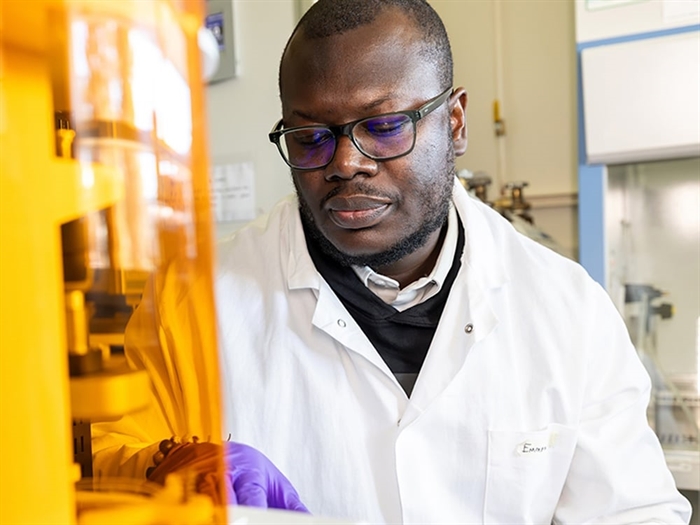
Osei is an assistant professor at the Irving K. Barber Faculty of Science, department of biology, at the University of British Columbia, and adjunct professor at the Centre for Heart Lung Innovation at UBC and St. Paul’s Hospital.
He oversaw the study at his lab, which was led by his students Sakshi Phogat and Tony Ju Feng Guo, who are co-first authors of the study.
3D bioprinted tissue is called a model. Creating a model that had three different types of cells in it and had a structure that mimicked blood vessels was very exciting because it’s not done much, Osei said.
Bringing cigarettes in was a way to stress test the model and prove that it was working as it should, Osei added.
Moving from 2D to 3D models
Osei said humans first figured out they could take cells out of the body and grow them independently a little over 100 years ago.
Cells from a heart, brain or lung would be grown on a flat surface, so they were considered 2D, with width and length, and could act as a model to learn how different cells reacted to things like drugs, disease or other factors in a controlled environment, he said.
But, he added, the human body isn’t 2D; it’s 3D, so drugs and treatments that seem to work in a 2D model rarely work in humans. Testing on animals also doesn’t always translate to use in humans because we’re different animals, he added.
Which is why, over the past decade or so, researchers have been working towards building 3D models. If those models are bioprinted, they could be built the exact same way every time — which is important for research — and include multiple types of cells, which is more similar to the complexity of actual human tissue.
To 3D print tissue, a researcher needs to tell the printer exactly where to put everything — which type of cell goes where, in what structure, and how many cells go in what order.
If that sounds complicated, it’s only scratching the surface of what’s going on, Osei said with a laugh.
It’s important to note that bioprinting doesn’t create cells. This study got its cells from various scientific sources, like cells built out of blood or tissue cells, or cancerous cells stored in labs, and then gave them to the printer.
The model used fibroblast cells to mimic the cells that create tissue structure, epithelial cells to mimic the cells that line the airway and endothelial cells inside of channels to mimic blood vessels.
The cells are laid out in a structure made of gelatin, a type of collagen that makes up about 70 per cent of the human body, and some other molecules, Osei said.
The finished model is pretty small — about 500 millimetres cubed, which is slightly smaller than an eight-millimetre dice, or comparable to a fingernail.
But that’s huge compared with the size of tissue samples researchers usually get, Osei said. When a doctor takes a tissue sample from a patient, for example during a biopsy, they will try to take as little as possible, which means researchers usually get something between six and 20 millimetres cubed, which is smaller than half of a single lentil.
Not printing lungs for transplant (yet)
Osei said the model is not yet ready for human transplant but that bioprinting organs is a long-term goal of the industry.
Bioprinted organs could have a lot of benefits, Osei said. They could be built out of a person’s own cells, for example, which would increase the chances of the body accepting the organ and reduce the amount of drugs a person has to take after a transplant.
In general, when a person receives an organ transplant, they will have to be on medications to suppress their immune system for the rest of their life so their body doesn’t attack the donated organ.
A near-term goal is improving researchers’ understanding of how lungs work and what’s happening when they don’t work, like when someone has an incurable disease such as chronic obstructive pulmonary disease or asthma, he said.
Osei said that in his PhD he showed that when cells don’t communicate properly it can lead to disease. Building 3D models will help him study how those communications are getting mixed up and how to better design drugs that address the root of the problem, rather than the symptoms.
Smoke ’em if you print ’em
It’s exciting to see that Osei’s model acted like human lung tissue when exposed to cigarette smoke, said Stephanie Willerth, a professor in biomedical engineering and director of the nanotherapeutics cluster at the department of mechanical engineering and division of medical sciences at the University of Victoria, and affiliate professor in the school of biomedical engineering at UBC.
Willerth was not involved in the study but also works on bioprinting tissue.
“It’s easy to print tissue but harder to get multiple cell types growing and working together,” Willerth said, adding that most 3D-printed models are still working with one or two cell types, so it was great to see three cells used in this model.
Vancouver is a hotbed for biotech companies. Aspect Biosystems, which has already printed a human kneecap, raised $150 million earlier this year to work on cures for serious diseases.
Osei said his lab has another project in the works that looks at how wildfire smoke affects the lungs.
“We’re in the Okanagan and wildfires are very important here,” he said.
— This article was originally published by The Tyee
News from © iNFOnews.ca, . All rights reserved.
This material may not be published, broadcast, rewritten or redistributed.

This site is protected by reCAPTCHA and the Google Privacy Policy and Terms of Service apply.
Want to share your thoughts, add context, or connect with others in your community?
You must be logged in to post a comment.